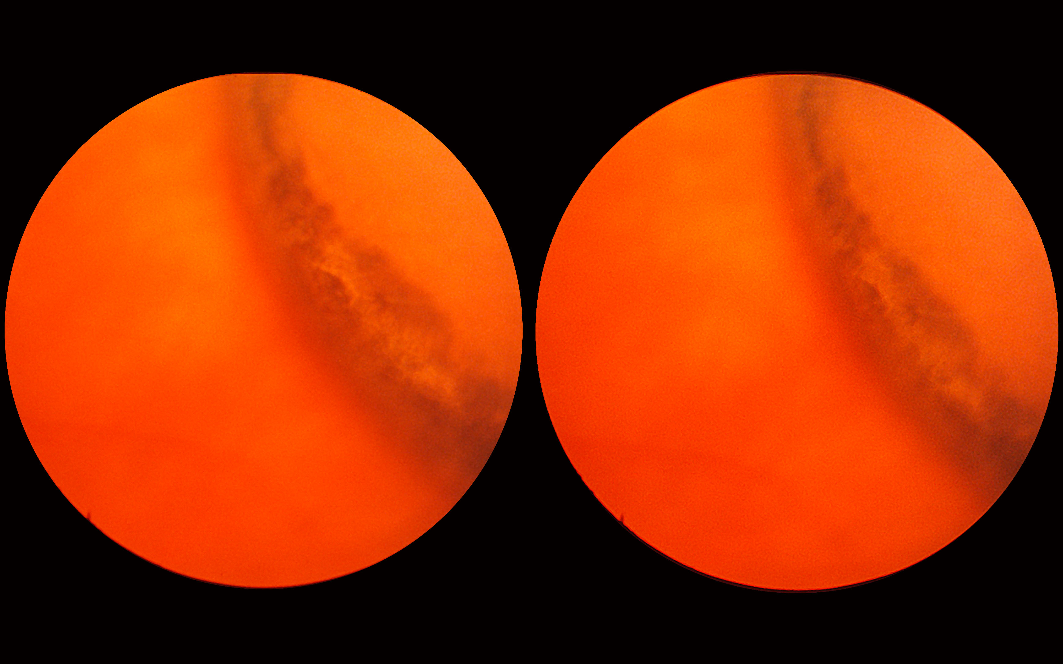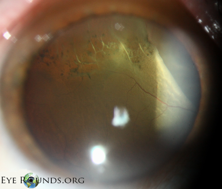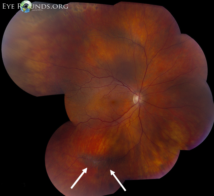

Approximately 18.7–29.7% of RRD is associated with lattice degeneration ( 1, 4). The prevalence of lattice degeneration is about 8% in the general population, and 16.9% in myopic patients ( 2, 3).

Lattice degeneration and retinal breaks are clinically significant peripheral retinal lesions that predispose patients to the development of rhegmatogenous retinal detachment (RRD) ( 1, 2). Keywords: Deep learning fundus image lattice degeneration retinal breaks ultra-widefield This system may help to prevent the development of rhegmatogenous retinal detachment by early detection of NPRLs. In the test set, 150 of 154 true-positive cases (97.4%) displayed heatmap visualization in the NPRL regions.Ĭonclusions: A DL system has high accuracy in identifying NPRLs based on UWF images. The best algorithm in each preprocessing method was InceptionResNetV2 (average AUC =0.996). The best preprocessing method in each algorithm was the application of original image augmentation (average AUC =0.996). Results: In the test set, the best DL system for identifying NPRLs achieved an area under the curve (AUC) of 0.999 with a sensitivity and specificity of 98.7% and 99.2%, respectively. Heatmaps were generated to visualize the process of the best DL system in the identification of NPRLs. An independent test set of 750 images was applied to verify the performance of 12 DL models trained using 4 different DL algorithms (InceptionResNetV2, InceptionV3, ResNet50, and VGG16) with 3 preprocessing techniques (original, augmented, and histogram-equalized images). The reference standard was determined when an agreement was achieved among all 3 ophthalmologists, or adjudicated by another retinal specialist if disagreements existed. All images were classified by 3 experienced ophthalmologists. Methods: A total of 5,606 UWF images from 2,566 participants were used to train and verify a DL system. Therefore, we aimed to develop and evaluate a deep learning (DL) system for automated identifying NPRLs based on ultra-widefield fundus (UWF) images. However, screening NPRLs is time-consuming and labor-intensive.

#These authors contributed equally to this work.īackground: Lattice degeneration and/or retinal breaks, defined as notable peripheral retinal lesions (NPRLs), are prone to evolving into rhegmatogenous retinal detachment which can cause severe visual loss. Zhongwen Li 1#, Chong Guo 1#, Danyao Nie 2#, Duoru Lin 1, Yi Zhu 1,3, Chuan Chen 1,3, Li Zhang 1, Fabao Xu 1, Chenjin Jin 1, Xiayin Zhang 1, Hui Xiao 1, Kai Zhang 1,4, Lanqin Zhao 1, Shanshan Yu 1, Guoming Zhang 2, Jiantao Wang 2, Haotian Lin 1ġState Key Laboratory of Ophthalmology, Zhongshan Ophthalmic Center, Sun Yat-sen University, Guangzhou 510060, China 2Shenzhen Ophthalmic Center, Jinan University, Shenzhen 518001, China 3Department of Molecular and Cellular Pharmacology, University of Miami Miller School of Medicine, Miami, Florida, USA 4School of Computer Science and Technology, Xidian University, Xi’an 710071, ChinaĬontributions: (I) Conception and design: Z Li, C Guo, D Nie, J Wang, H Lin (II) Administrative support: H Lin (III) Provision of study materials or patients: J Wang, G Zhang, D Nie (IV) Collection and assembly of data: Z Li, D Lin, L Zhang, F Xu, X Zhang, H Xiao, L Zhao, S Yu (V) Data analysis and interpretation: Z Li, C Guo, D Nie, L Zhang, F Xu, H Lin, D Lin, Y Zhu, C Chen, C Jin, K Zhang, J Wang, G Zhang, H Xiao, L Zhao (VI) Manuscript writing: All authors (VII) Final approval of manuscript: All authors. Policy of Dealing with Allegations of Research Misconduct.

Policy of Screening for Plagiarism Process.


 0 kommentar(er)
